
Fig. 4.
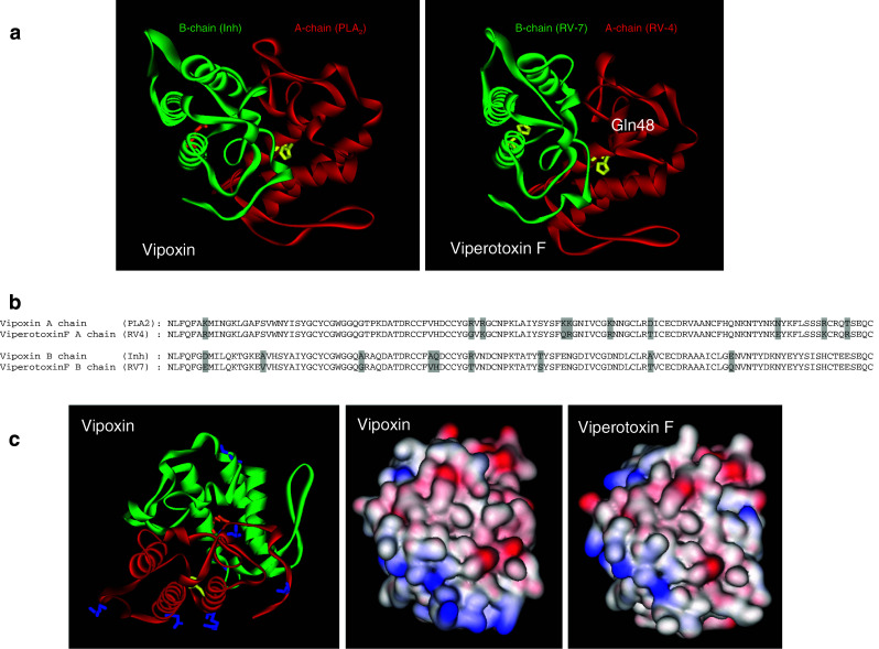
a Ribbon model of vipoxin (PDB ID: 1AOK) and viperotoxin F (PDB ID: 1OQS) showing PLA2 subunit (red) and its inhibitor (green). The active site residue, His48, is replaced with Gln48 in the inhibitor, which is shown in a stick and ball model. b Alignment of amino acid sequence of chain A and B of vipoxin and viperotoxin F. The substituted amino acid residues are highlighted in grey. c The surface residues that are substituted in vipoxin are shown in a stick model (blue color). The surface charge of vipoxin and viperotoxin F is shown