Fig. 6.
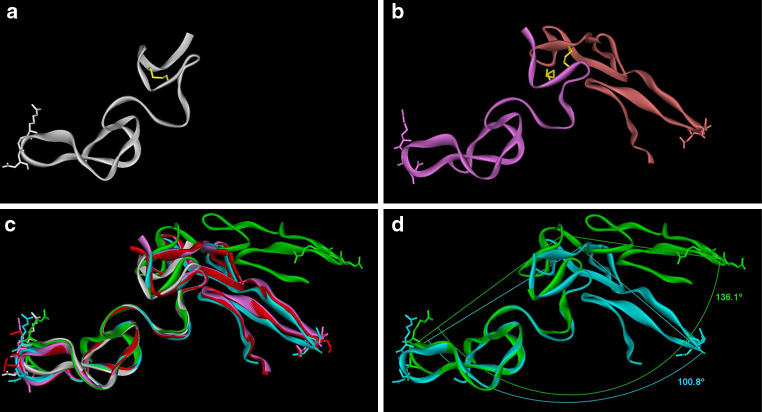
Dimeric disintegrins. (a) Disintegrin monomer (PDB ID: 1J2L) (white ribbon). RGD is shown as a white stick model. Intramolecular disulfide bond (C6:C14) is shown in the yellow stick. b Homodimeric disinegrin (PDB ID: 1RMR) (pink and orange ribbons). Side chains of RGD are shown as pink/orange sticks. Intermolecular disulfide bonds (C7:C12) are shown in the yellow stick. c Superimposition of homodimeric disintegrins (PDB ID: 1RMR in pink ribbon, PDB ID: 1TEJ in cyan ribbon) and heterodimeric integrins (PDB ID: 1Z1X in red ribbon, PDB ID: 3C05 in green ribbon) on monomeric disintegrin (PDB ID: 1J2L in white ribbon). RGDs are shown as the white stick in 1J2L, as the pink stick in 1RMR, as the cyan stick in 1TEJ and as the green stick in 3C05. MLD is shown as the red stick in 1Z1X. d For 1RMR, the angle between chain A Gly43 Cα, chain B Cys6 Cα, and chain B Gly43 Cα is 100.8°; for 1Z1X, the angle between chain A Gly44 Cα, chain B Cys7 Cα and chain B Gly44 Cα is 136.1°

