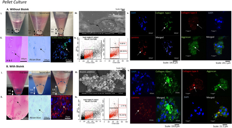
Figure 3.
Biomimetic pellet tissue construct characterisation for BMSCs differentiated chondrocytes. The figure shows the BMSCs-induced chondrocytes characterisation (A) without bioink (i) cultured and monitored for increasing pellet size for three weeks (ii) H&E staining showed oval-shaped morphology with alcian blue demonstrated GAG accumulation around the cells and CLSM imaging showed BMSCs differentiation marker for chondrogenesis VCAM1 positive expression. (iii) SEM imaging showed fewer cells growing individually without bioink (iv) Cell viability analysis with 7-AAD dye using flow cytometry showed R1 as the desired population, R2 as the dead cell population, and R3 as the live cell population showing the percentage viability of the cultured construct. (v)The ECM-specific markers showed positive expression for collagen type I, aggrecan (Alexa Fluor 488 conjugated) and laminin and minimal expression for collagen type II (Alexa Fluor 594 conjugated). (B) With bioink (i) the pellet increased in size to 0.8 cm after three weeks of culture. (ii) H&E staining revealed isogenous clusters of cells, increased GAG content and increased expression of VCAM 1 in proliferating BMSCs-induced chondrocytes. (iii) The SEM imaging showed cellular attachment on bioink additive (iv) The 7AAD viability assay showed an increase in the percentage of the live cell population R3. (v) CLSM characterisation revealed an increase in chondrogenesis secreted markers collagen type II expression, in addition to collagen type I, aggrecan with cell-ECM marker laminin expression, and counterstaining with the nuclear dye DAPI (blue).

