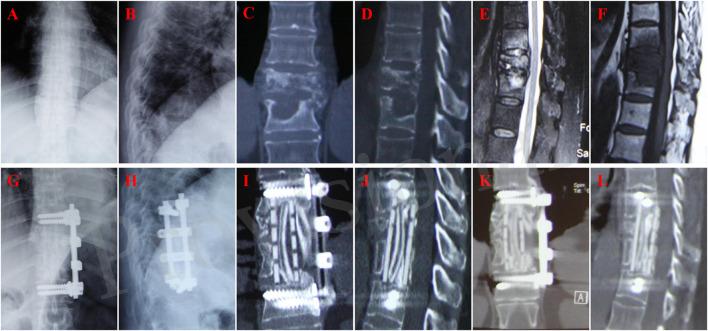
FIGURE 2.
A 60-year-old female with T10-11 tuberculosis in n-HA/PA66 group and received one-stage lesion debridement, n-HA/PA66 composite cage placement, allogeneic bone interbody fusion, and instrumentation. (A–F) Preoperative X-ray, 3D CT and MRI showing destruction of T10-11 intervertebral disc and adjacent vertebral bodies. (G–H) X-ray immediate after surgery. (I, J) 3D CT at 12 months after surgery showing good internal fixation position. (K, L) 3D CT at 46 months after surgery showing solid bone fusion and no significant subsidence of the graft.

