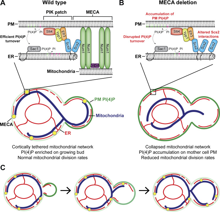
Figure 10.
The mitochondria–ER–PM contact site mediated by Num1 regulates the distribution of PI(4)P and mitochondrial dynamics. (A) MECA forms a tripartite contact site that tethers mitochondria to the PM and ER. Under wild type circumstances, PIK patches localize adjacent to MECA contact sites and synthesize PI(4)P (depicted as green lipids) from the precursor PI (depicted as magenta lipids). PI(4)P is then presented to the ER-localized Sac1 phosphatase where it is hydrolyzed into PI. Wild type levels of PI(4)P synthesis and turnover result in a robust enrichment of PI(4)P on the PM of the growing bud. (B) Loss of MECA results in a collapse of the mitochondrial network and PI(4)P accumulation on the mother cell PM. Loss of the Num1–Scs2 interaction may alter PI(4)P distribution by changing the amount of Scs2 available to interact with FFAT motif containing proteins. Additionally, loss of MECA may disrupt PI(4)P turnover by preventing PI(4)P from being properly presented to Sac1. Loss of MECA also severely perturbs mitochondrial division rates. (C) Changes in PM PI(4)P levels coincide with the formation of new mitochondria–ER–PM contact sites. Newly formed buds contain little cortical ER, have yet to inherit mitochondria, and maintain high levels of PM PI(4)P. As the cell cycle progresses, more cortical ER as well as mitochondria are inherited, MECA contact sites form, and bud PM PI(4)P levels begin to decrease. When the cell is near cytokinesis, the cortical ER and mitochondrial network are stably anchored via MECA and other MCSs, and the amount of PI(4)P on the bud PM decreases to a level that is comparable to the mother cell PM. The black arrows are used to indicate progression through the cell cycle. Darker shades of green represent higher concentrations of PM PI(4)P.

