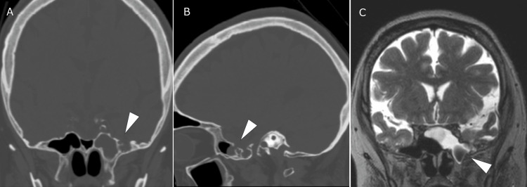
Figure 1. The CT and MRI scans show meningoencephalocele protruding into the left sphenoid sinus through the bone defect and CSF in the sphenoid sinus.
Head CT showing a bone defect (arrowheads, A and B) in the left middle cranial fossa communicating with the sphenoid sinus. MRI showing meningoencephalocele in the sphenoid sinus (arrowhead, C) and accumulated cerebrospinal fluid within the sphenoid sinus.
CT: computed tomography; MRI: magnetic resonance imaging; CSF: cerebrospinal fluid

