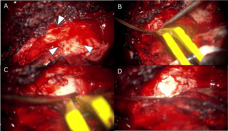
Figure 2. A left frontotemporal craniotomy was performed, and meningoencephalocele was excised approaching the extra dural side.
Multiple bone pores (arrowheads, A) are observed, possibly related to thinning of the middle cranial fossa. Brain tissue is observed deviating from the bone defect and is cauterized (B and C). Repair of the bone defect using surgical bone cement (D).

