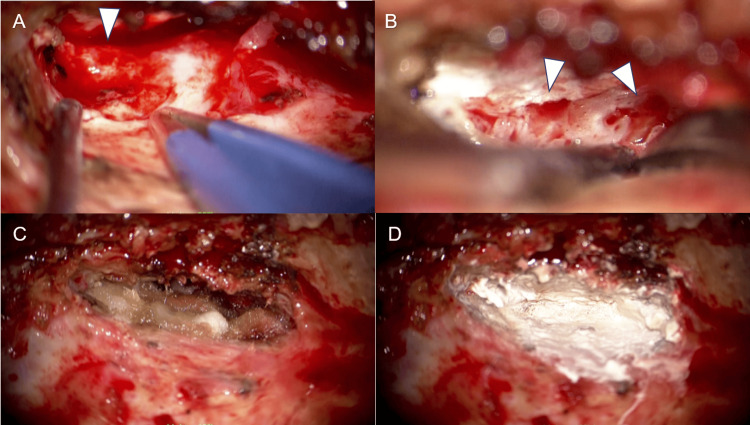
Figure 3. During the second surgery, residual bone defects and surrounding bone pores were observed and closed.
The previously identified dural defect remained open, allowing the leakage of CSF (A). There were bony pores around the leakage point (arrowheads, B). The dural defect was closed using temporal fascia and fibrin glue (C). The middle cranial fossa was extensively repaired with bone cement while also covering the surrounding bone pores (D).
CSF: cerebrospinal fluid.

