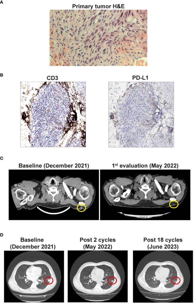
Figure 1.
(A) Primary tumor histology. Hematoxilyn-eosin (H&E) staining of the primary tumor. (B) Left panel: Non-brisk T cell infiltration (anti-CD3 immunohistochemistry). Right panel: Negative tumor PD-L1 expression. Magnification 20x. (C) CT scan showing the cutaneous and subcutaneous lesions (yellow circles) before the start of the therapy (Baseline) and after 4 cycles (1st evaluation). (D) CT scan of the lung metastases (red circles) before the start of the therapy (Baseline), after 3 cycles, and after 18 cycles of immunotherapy.

