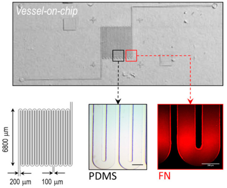
Figure 4.
Microfluidic vessel-on-a-chip model. (Upper) Optical microscopy image of the polydimethylsiloxane microfluidic chip. (Lower) Left—dimensions of the microchannel; middle and right—magnified image of the fabricated and fibronectin-coated (rhodamine) channel. Reproduced under the Creative Common CC BY 4.0 license Reprinted from Ref. [24].

