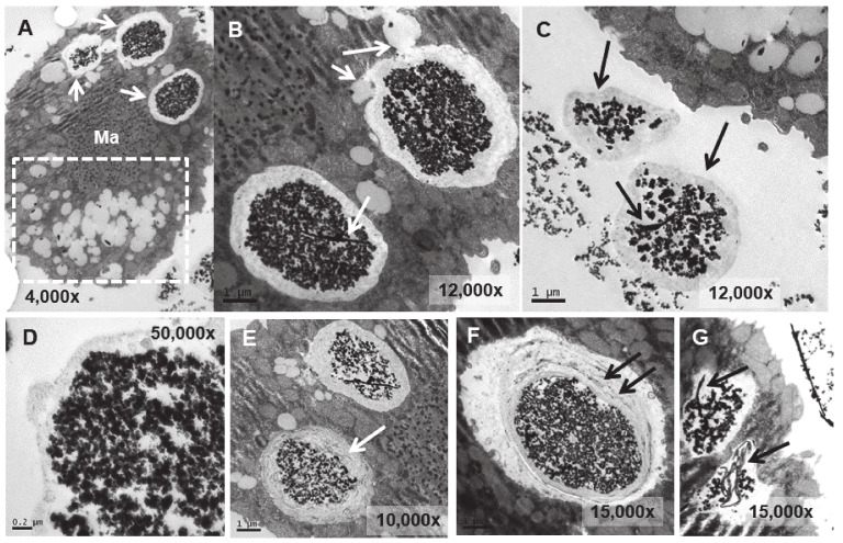
Figure 7.
TEM images of TtEuCl3-adap cells. (A): Vacuoles with electrodense condensed granular content and surrounded by an electrolucent material originating from fusion with numerous lipid droplets (arrows). Region with numerous lipid droplets (dashed line box). Ma (macronucleus). (B): Magnified region from panel (A). (C): The granular contents from vacuoles (arrows) with their peripheral electrolucid region are excreted out of the cell. (D): Magnified detail of one of the excreted materials. (E,F): The electrolucent material sometimes contains membranous or fibrillar elements surrounding the electrodense granular content (arrows). (G): In some vacuoles, an electrodense fibrillar content is observed (arrows) together with the granular one.

