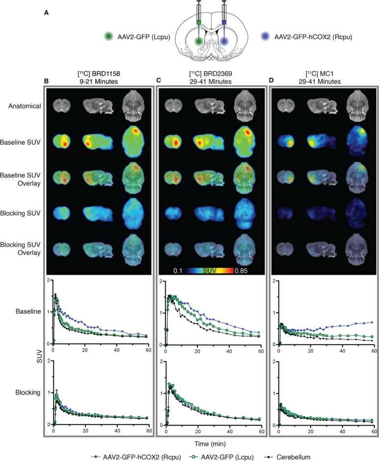
Figure 2.
Brain PET intrasubject comparison of localized COX-2 overexpression (intrastriatal AAV2-GFP-hCOX2) with three different COX-2 radiotracers in one rat (animal ID: SD2006071) across three imaging sessions. [11C]BRD1158 is an effective COX-2 PET radiotracer that demonstrates uniquely fast onset in a rodent COX-2 overexpression model. (A) Schematic of injection paradigm in rats showing ICV injection of AAV2-GFP-hCOX2 (6.53 × 10¹²gc/mL) to right caudate (in blue), to induce overexpression of COX-2 and AAV2-GFP to left caudate (in green), as control. Animals were also given IP mannitol (10 mL/kg dose) to enhance transgene expression and increase vector spread. (B–D) Time average SUV images (above) and regional time activity curves (below) in one rat using tracer [11C]BRD1158 at 57 days post-AAV injection (left, B), [11C]BRD2369 at 61 days post-AAV injection (middle, C), and [11C]MC1 at 75 days post-AAV injection (right, D), at baseline and celecoxib (1 mg/kg) blocked conditions. Time averages shown are from the optimal portion of the scan; 9–21 min for [11C]BRD1158, and 29–41 min for [11C]BRD2369 and [11C]MC1. Representative MRs are shown. (See Figure S8 for a technical replicate in a second rat.)

