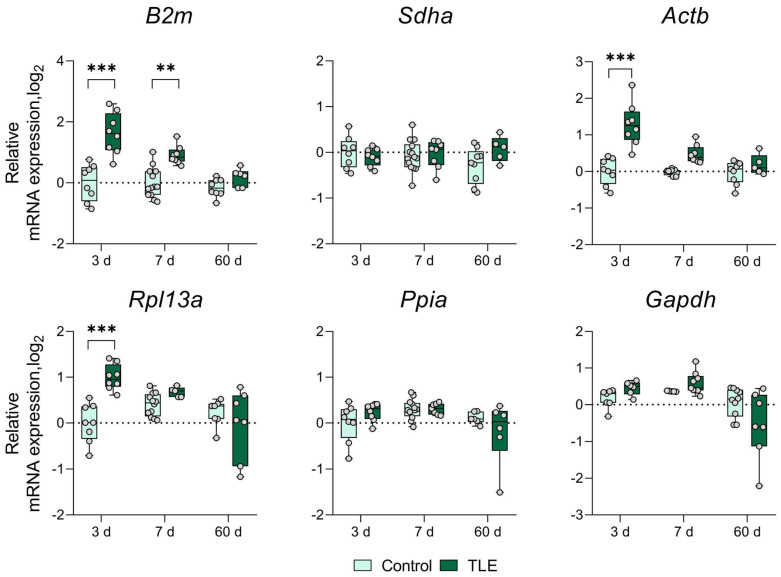
Figure A2.
Changes in the expression of commonly used reference genes in the temporal cortex area. Analysis was performed 3, 7, and 60 days after pilocarpine-induced status epilepticus. The data were normalized to the expression levels of the three most stable genes (Ywhaz, Hprt1, and Pgk1) in the rat lithium–pilocarpine model of temporal lobe epilepsy. **, *** p < 0.01 or p < 0.001, respectively (two-way ANOVA followed by Sidak post hoc test); Control—control group, TLE—experimental group. All the data are presented as individual values (circles) with the minimum, maximum, sample median, and first and third quartiles.

