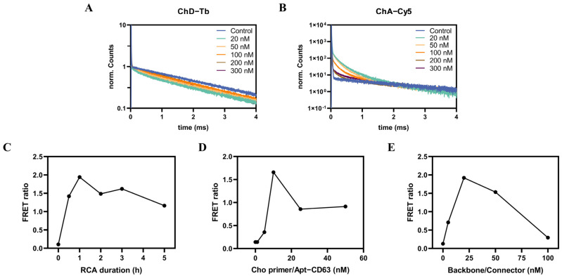
Figure 4.
The optimization of the experimental conditions. The normalized fluorescence decay curves of the donor (A) and acceptor channels (B) corresponding to various concentrations (ranging from 20 nM to 300 nM) of Tb probe/Cy5 probe are shown. The fluorescence decay curves of all control measurements (without the presence of SH−SY5Y exosomes) in the donor and acceptor channel were normalized to be the same. Tb quenching and Cy5 sensitization in the presence of SH−SY5Y exosomes compared to that of control measurements demonstrated the FRET from Tb to Cy5. Also shown are the effects of the RCA duration (C), PLA probes (D), and backbone/connector (E) on FRET ratios. All measurements were performed with SH−SY5Y exosomes (20 ng∙μL−1), Cho primer/Apt−CD63 (10 nM), backbone/connector (10 nM), and Tb probe/Cy5 probe (20 nM) and the RCA duration time was set to 2 h, except in the special conditions indicated in the figures.

