Figure 4.
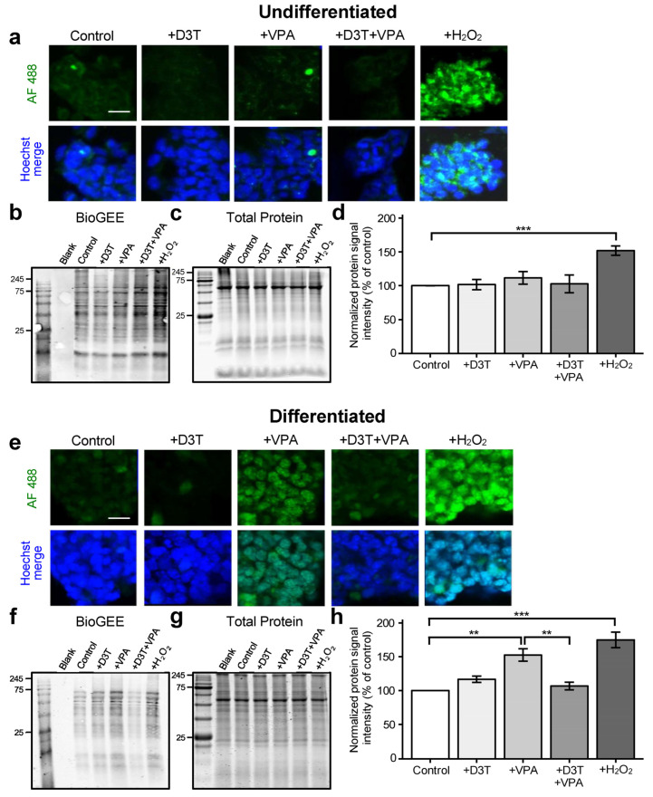
Protein S-glutathionylation is unchanged in undifferentiated cells but increased in differentiated neurons following VPA exposure. Undifferentiated cells and differentiated neurons were treated and collected for protein S-glutathionylation detection using biotinylated glutathione ethyl ester (BioGEE), a GSH analog. (a) Undifferentiated cells were stained with a streptavidin-conjugated fluorophore, Alexa Fluor 488 (AF 488), to detect BioGEE and then imaged using confocal microscopy. (b–d) Undifferentiated cells were assessed for BioGEE signal intensity (b), normalized to their respective GelCode Blue-stained samples run in parallel (c), and analyzed using the normalized protein signal intensity (n = 3; (d)). (e) Differentiated cells were similarly probed with BioGEE and imaged. (f–h) Differentiated neurons were assessed for BioGEE signal intensity (d), normalized to their respective GelCode Blue-stained samples (g), and analyzed using the normalized protein signal intensity (n = 3; (h)). Normalized BioGEE signal intensity is a direct measure of protein S- glutathionylation. Brief exposure to H2O2 was used as a positive control (a–h). Scale bars represent 50 μm (a,e). Images are representative of three independent experiments (a,e) and data are presented as means ± SEM (d,h). Statistical comparisons were made using a one-way ANOVA followed by a pairwise t-test using the Bonferroni correction (d,h). Asterisks denote a statistically significant difference (** = p < 0.01, and *** = p < 0.001; (d,h)).

