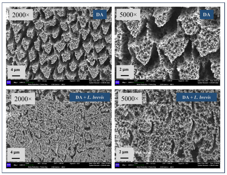
Figure 2.
Representative SEM micrographs of specimens at 2000× and 5000× magnifications. The top micrographs (specimens treated with DA) highlight a complete loss of surface integrity, with firm prism core dissolution and loss of interprismatic rods protruding, along with damaged and fragmented enamel. At increased magnification (5000×), pits, pores, and micro-erosion were well observed. The bottom micrographs (specimens treated with DA + L. brevis) display a reduced dissolution of the prisms and fewer holes and eroded areas than the specimens treated with the demineralizer alone.

