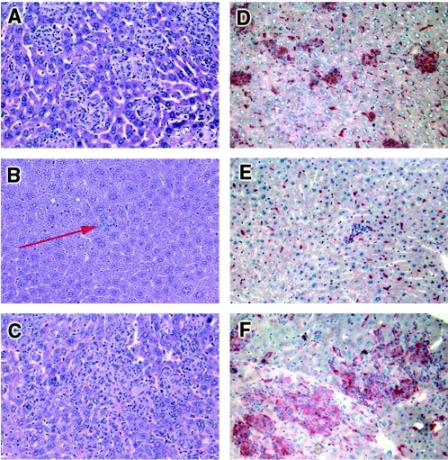
FIG. 2.
Impaired granuloma formation and reduced macrophage activation in transgenic mice expressing high levels of sTNFR1 and enhanced macrophage activity in transgenic mice expressing low levels of sTNFR1 after 2 weeks of BCG infection. Histological tissue sections were obtained from livers after 2 weeks of BCG infection. Hematoxylin and eosin staining revealed well-defined granulomas in control mice (A), small granuloma-like structures in transgenic mice expressing high levels of sTNFR1 (arrow) (B), and large granulomas in transgenic mice expressing low levels of sTNFR1 (C). (D) Strong staining for acid phosphatase activity (marker of macrophage activation) in control granulomas. (E) Absence of activity in granulomas from transgenic mice expressing high levels of sTNFR1. (F) Increased staining in the large granulomas from transgenic mice expressing low levels of sTNFR1. The results are representative of the results of two independent experiments (five mice per group). Magnification, ×200.

