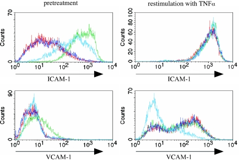
FIG. 4.
Phenotype of PRBC-stimulated HUVEC after contact with TNF-α. HUVEC were incubated for 20 h with medium (red line), RBC alone (dark blue line), TNF-α (10 ng/ml) (green line), or PRBC (light blue line), and cells were washed three times with medium and cultivated for an additional 6 h with medium or with TNF-α (10 ng/ml). Cells were analyzed by flow cytometry after indirect immunostaining with antibodies recognizing ICAM-1, VCAM-1, and E-selectin. The histograms show the expression levels of the surface molecules.

