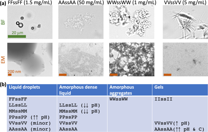
Fig. 2.
Four forms of condensates formed by tetrapeptides. (a) Contrasting the four forms of condensates by brightfield (BF) and negative-stain electron microscopy (EM). Samples were prepared at the indicated concentrations in milli Q water and pH 13. The green scale bar applies to all the BF images; all the brown scale bars represent 200 nm. (b) Summary of tetrapeptides that form each form of condensate. Single arrows mean roughly one-half of the phase-separation pH range; double arrows mean a small fraction of the phase-separation pH or concentration range.

