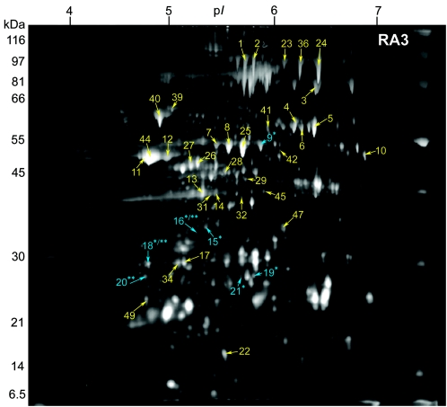
FIG. 1.
2-DE gel of the secreted proteins of B. anthracis RA3 (pXO1+ pXO2+) grown under inducing conditions (0.85% [wt/vol] bicarbonate, 5% CO2, 37°C) in minimal R medium. Samples containing 100 μg of TCA-precipitated extracellular proteins were focused on pH 4 to 7 IPG strips and separated on 10% Duracryl gels. The gels were stained with SYPRO Ruby fluorescent stain and imaged at 470 nm. Of the 42 identified spots, three (blue; **) are unique to RA3 compared to RA3R; six spots (blue; *) are exclusive to RA3 relative to RA3:00; two spots (*/**) are unique to RA3 compared to both RA3R and RA3:00. Molecular masses are indicated on the left.

