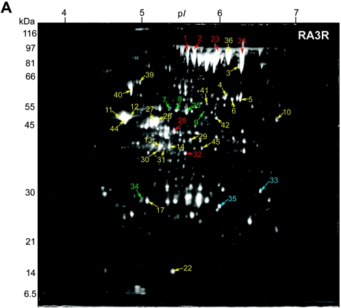
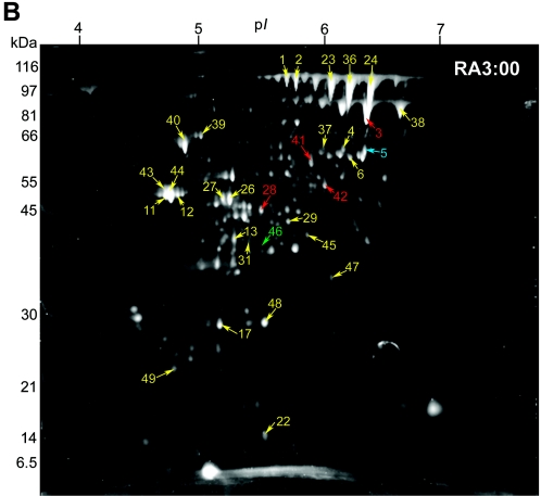
FIG. 2.
Identified secreted-protein spots in (A) B. anthracis RA3R (pXO1+ pXO2−) and (B) B. anthracis RA3:00 (pXO1− pXO2−), both grown under inducing conditions (0.85% [wt/vol] bicarbonate, 5% CO2, 37°C) in minimal R medium. Secretome samples containing 100 μg total protein were focused in the first dimension on pH 4 to 7 IPG strips and separated in the second dimension using 10% Duracryl gels. The gels were then stained with SYPRO Ruby fluorescent stain and imaged at 470 nm. Unique, up-regulated, and down-regulated protein spots in RA3R or RA3:00 compared to RA3 are labeled in blue, red, and green, respectively. Protein spots observed at similar expression levels (constitutive) in RA3R or RA3:00 relative to RA3 are labeled in yellow. Molecular masses are indicated on the left.


