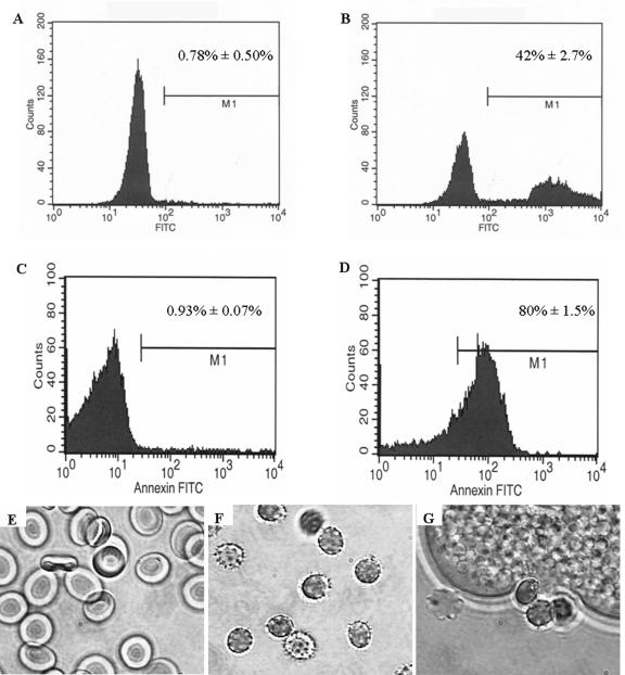
FIG. 2.
Erythrocytes exposed to Entamoeba histolytica redistribute phosphatidylserine to the outer leaflet of the membrane and undergo morphological changes. (A to D) PS exposure was measured using annexin V-FITC staining of erythrocytes. Representative histograms of healthy erythrocytes and erythrocytes incubated with E. histolytica trophozoites are shown in panels A and B, respectively. Similarly, annexin V-FITC staining was performed on erythrocytes exposed to treatment with 2.5 mM calcium. Representative histograms of healthy erythrocytes in HEPES buffer for 48 h and erythrocytes in HEPES buffer exposed to 2.5 mM CaCl2 are shown in panels C and D, respectively. (E to G) Representative micrographs of healthy erythrocytes (panel E), crenulated erythrocytes following 48 h calcium treatment (panel F), and erythrocytes following 20 min exposure to E. histolytica (panel G).

