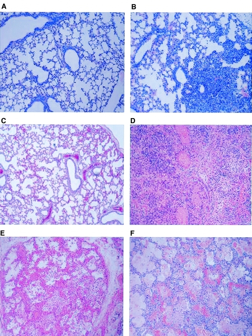
FIG. 3.
Histopathology. (A and B) Photomicrographs of lung biopsies taken from a 21-day-old uninfected BALB/c mouse (A) and a BALB/c mouse infected with 5 × 10⁸ CFU B. pertussis by aerosol (B) 10 days after challenge, obtained with a 10× objective plus digital zoom. (C and D) Photomicrographs of lung biopsies taken from newborn piglets 4 days after treatment with 5 × 10⁹ CFU heat-inactivated B. pertussis (4× objective plus digital zoom) (C) or after challenge with 5 × 10⁹ CFU B. pertussis (10× objective plus digital zoom) (D). (E and F) Photomicrographs of lung biopsies taken from two children who died at 29 (E) or 37 (F) days of age after infection with B. pertussis (10 × objective plus digital zoom).

