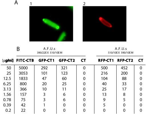
FIG. 4.
Analysis of CT chimera fluorescence. (A) Expression of the GFP-CT and RFP-CT chimeras in E. coli after an overnight incubation with 0.2% l-arabinose: E. coli NovaBlue plus pTatABCE plus pJKT35 with FITC filter (frame 1) and NovaBlue plus pTatABCE plus pJKT36 with rhodamine filter (frame 2). Both fluorescent proteins exhibit polar localization, and diffuse fluorescence of the periplasm is also evident from the mRFP-CT chimera. (B) Fluorescence spectroscopy of the purified GFP-CT and mRFP-CT chimeras. The numbers 1 and 2 in the designations indicate different purified preparations of the same chimera.

