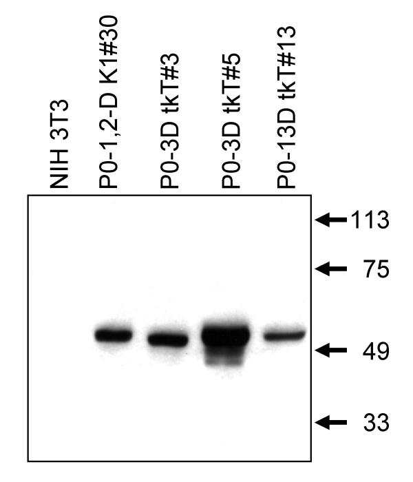
Figure 1.
GFAP expression in the Linker tkT cell lines. The tkT cell lines and NIH 3T3 cells were grown in 1 mM dibutyryl cAMP for 3 to 8 days. Intermediate filament extracts from 1 to 6 × 10⁵ cells were loaded in each lane and subjected to SDS PAGE and Western blotting. The membranes were immunostained for GFAP, which is specific for astrocytes. All of the astrocyte cell lines expressed GFAP and the NIH 3T3 cells did not.

