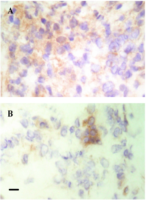
FIG. 2.
Representative microphotographs of tissue sections stained for TNF-α: cryosections of biopsies taken from a patient with T1R before prednisolone treatment was started (A) and after 1 month of prednisolone treatment (B). Bar = 10 μm. Harris hematoxylin counterstaining was used. Original magnification, ×400.

