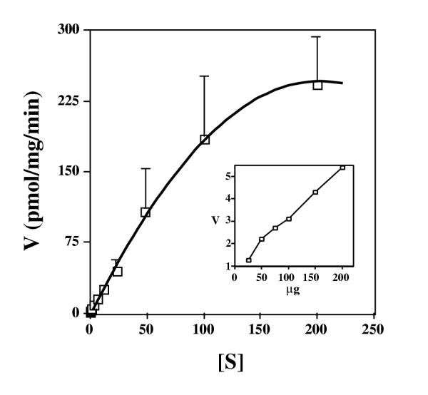
Figure 2.
The rate of uptake of ThDP by mitochondria isolated from normal lymphoblasts. Mitochondria were isolated from normal lymphoblasts and were incubated for 15 minutes with various concentrations of radioactive ThDP. Mitochondrial-associated counts were determined, and the velocity (V) (pmol ThDP per mg mitochondrial protein per min.) is plotted versus the concentration in micromolar of ThDP ([S]). Error bars represent SEM for four independent experiments. The inset shows uptake (V, pmol ThDP per mg mitochondrial protein per min.) versus varying amounts of resuspended mitochondria (μg of protein) in the presence of 2 M radioactive ThDP.

