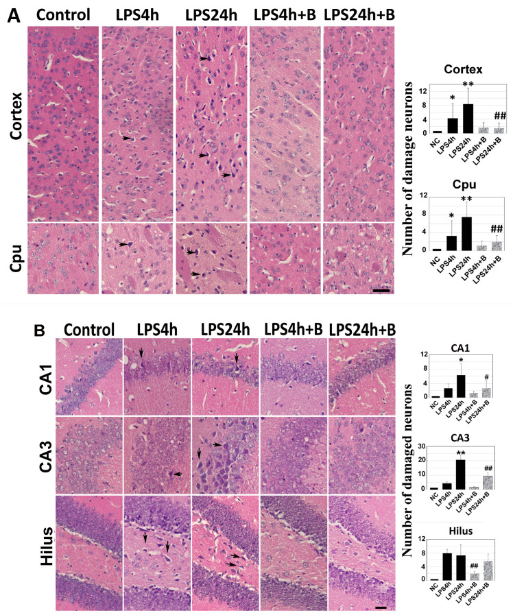
Figure 3.
Histologic outcomes in LPS and B355252 treated animals: (A) H&E staining in the cortex and caudate putamen (Cpu); (B) H&E staining in the hippocampal CA1, CA3, and hilus. LPS administration led to increased cell death at 24 h in the cortex, Cpu, CA1, CA3, and hilus, while treatment with B355252 ameliorated the damage. The data presented in the bar graphs represent the mean +/− SD. Significance levels are denoted as *, ** for p < 0.05, 0.01 vs. NC (naïve control); and #, ## for p < 0.05, 0.01 vs. the respective LPS counterpart. The scale bar represents 100 µm.

