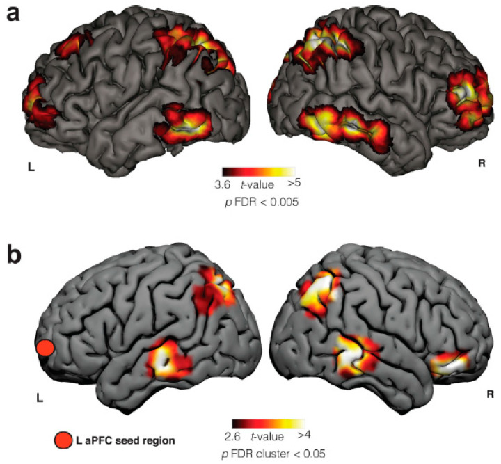
Figure 1.
Frontal, parietal, and temporal brain areas are involved in LD, which is evidenced by two different methods of neuroimaging: (a) Blood-oxygen-level-dependent (BOLD) activity in an fMRI case report of LD. (b) Seed-based resting-state (SBRS) functional connectivity differences between frequent lucid dreamers and non-frequent lucid dreamers (control group). Adapted from [15] with permission from the authors. R = right side and L = left side. aPFC = anterior prefrontal cortex.

