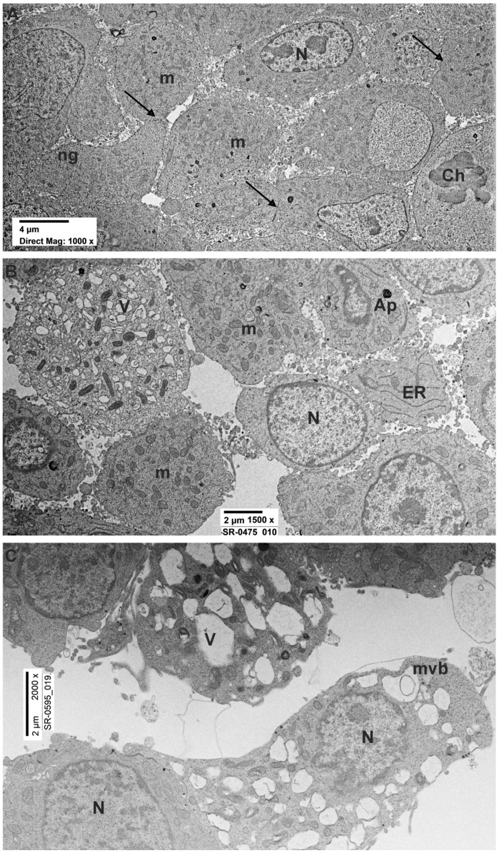
Figure 1.
Ultrastructural evaluation of untreated, rotenone 24–48 h PC12 cells. (A) Ultrastructure of control PC12s showing evident nuclei (N) delimited by a continuous nuclear membrane and numerous round/ovoid mitochondria (m). Intercellular contacts (arrows) were present among the cells. Numerous neuropeptides granules (ng) were visible. (B) Ultrastructure of PC12 treated with rotenone 0.5 µM for 24 h showing large nuclei delimited by an intact nuclear membrane and numerous mitochondria. Intercellular contacts among the cells were partially lost. Degenerating cells with several vacuoles (V) were present. (C) Representative TEM micrograph of PC12 treated with rotenone 0.5 µM for 48 h displaying clear signs of cell degeneration. Numerous vacuoles and isolated multivesicular bodies (mvb) were visible. Ch: chromosomes; ap: autophagosome; ER: endoplasmic reticulum.

