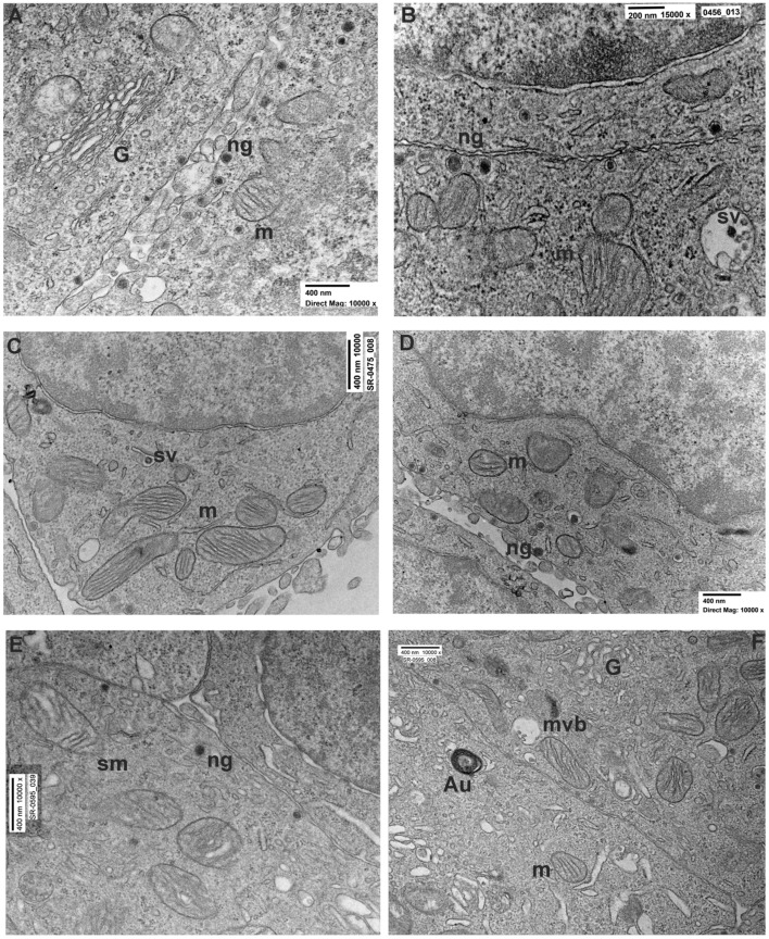
Figure 2.
Ultrastructural comparison of neuropeptide granules and synaptic vesicles in untreated and rotenone 24–48 h PC12 cells. (A,B) High magnification of two cell borders in the untreated PC12 group. Note the presence of numerous neuropeptides granules (ng) attached to the plasma membrane. Numerous Golgi cisternae (G) and mitochondria (m) with visible cristae were observed. (C,D) Representative picture of PC12s treated with rotenone 0.5 µM for 24 h showing the reduced presence of neuropeptide granules in proximity of the cell borders and elongated mitochondria with evident cristae. (E,F) Ultrastructure of PC12 treated with rotenone 0.5 µM for 48 h showing an evident reduction of neuropeptide granules, mitochondria with sign of swelling (sm), and multivesicular bodies (mvb) containing altered synaptic vesicle (sv).

