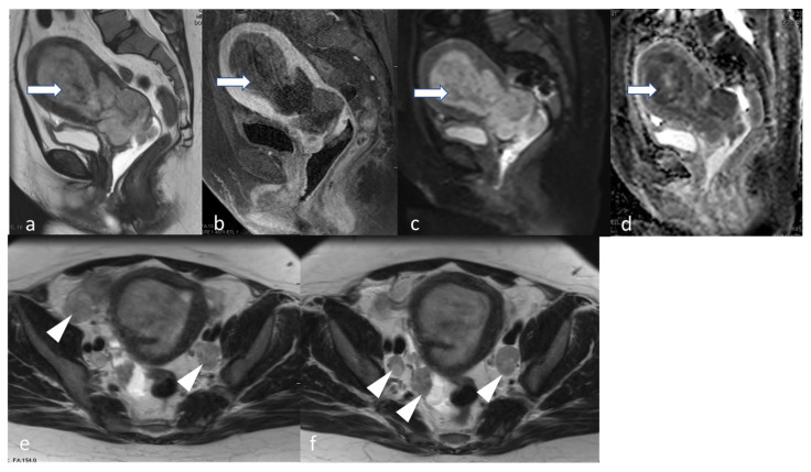
Figure 12.
Stage IIIC1ii. A 48-year-old female patient with high-grade endometrial carcinoma. Sagittal T2 (a) and T1 fat sat post-contrast (b) show a large mildly T2 hyperintense hypovascular endometrial mass (arrow) causing expansion of the endometrial cavity with diffusion restriction evidenced by a relatively high signal in DWI (c) and low ADC value (d). The mass extends inferiorly through the cervical canal involving the posterior lip and the posterior final fornix. Axial T2 images (e,f) show bilateral iliac metastatic lymph nodes (arrowhead).

