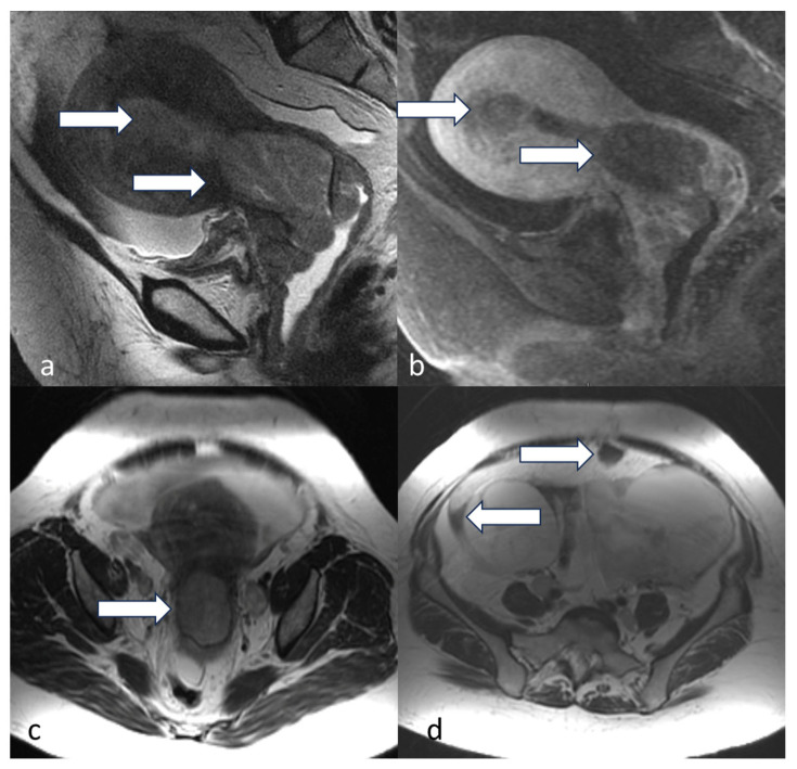
Figure 15.
Stage IVB. A 52-year-old female with serous endometrial carcinoma. Sagittal T2 (a), sagittal T1 post-contrast (b), and axial T2 (c) weighted MRI images show thickened irregular endometrium (arrow) with involvement of the cervix as well as the upper vagina. Axial (d) T2 weighted MRI image peritoneal involvement with moderate-volume ascites and peritoneal implant (arrow).

