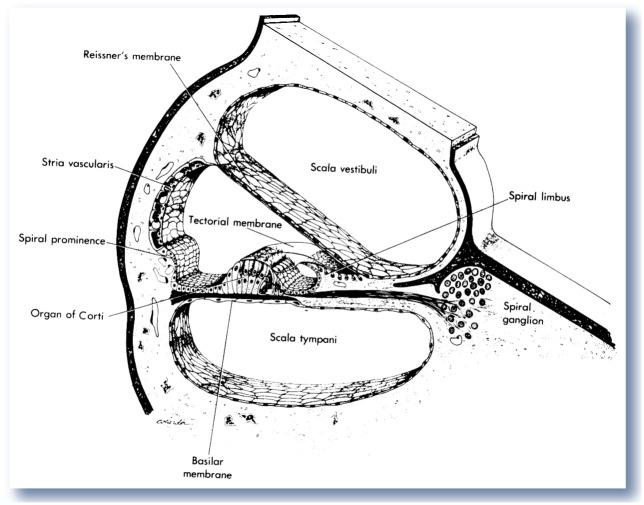
Figure 3.
Structures of the cochlea in cross-section. Inner hair cells in the organ of Corti are the primary sensory transduction cells, while the outer hair cells utilize active processes to increase or decrease sensitivity. Pigment cells in the stria vascularis play a major role in maintaining high potassium levels in the cochlear duct to support hair cell viability. Hair cells synapse on neurons of the spiral ganglion, which in turn become components of cranial nerve VIII. Reproduced from Bloom and Fawcett,8 with permission

