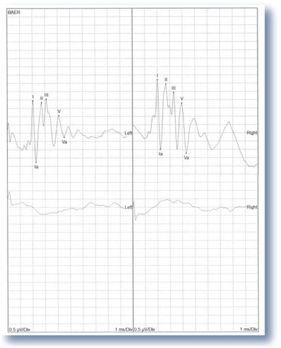
Figure 7.

(top) BAER recording from a 2-year-old normal cat, with peaks I, II, III and V labeled on the tracing. Peak I is generated by cranial nerve VIII entering the brain, while the later peaks are generated in the brainstem. (bottom) By comparison, this essentially flat line is a recording from a bilaterally deaf cat. 0.5 μv/div and 1 ms/div
