Figure 7.
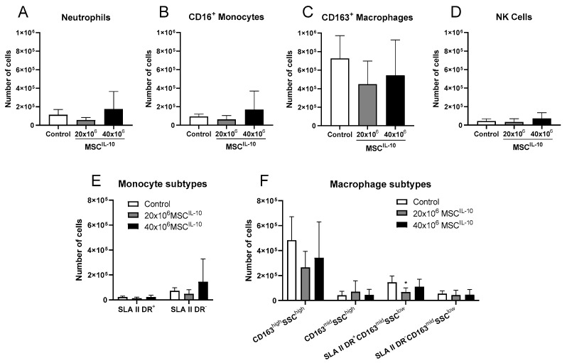
Lung transplant myeloid cell composition after MSCIL-10 treatment 3 days after transplantation. Myeloid cells and monocyte and macrophage subtypes in lung transplants 3 days after transplantation were quantified by flow cytometry. Lung (A) neutrophils, (B) CD16+ monocytes, (C) CD163+ macrophages and (D) NK cells. (E) Monocytes were further divided according to SLA II DR expression, and (F) macrophages according to CD163, side scatter and SLA II DR expression. Treatment with 20 × 106 MSCsIL-10 significantly decreased the number of SLAII-DR+CD163midSSClow-activated macrophages. Data mean ± standard deviation and analyzed by 1-way ANOVA with Dunnett’s correction comparing treatment groups to the control group. * p < 0.05. MSC, mesenchymal stromal cell; NK, natural killer; SLA, swine leukocyte antigen; SSC, side scatter.

