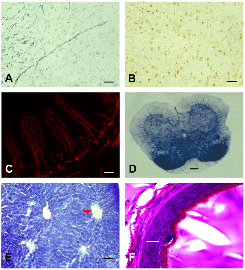
Figure 6.
Scalability of the gelatin embedding matrix to various tissue types in different mammalian species. (A,B) GFAP (astrocytes) brightfield immunohistochemistry was evaluated in rat (gray) and human (brown) brain cortical tissue. (C) Murine intestinal tissue sections demonstrated blood vessel (CD31; red) fluorescent detection. (D) Enzyme stains were used to evaluate alkaline phosphatase activity in murine spinal cord sections. (E,F) Standard histological Nissl and H/E were used to evaluate general morphology in the murine liver (red arrow = central vein) and human arterial vessel, respectively. Scale bar = 50 µm (A–F).

