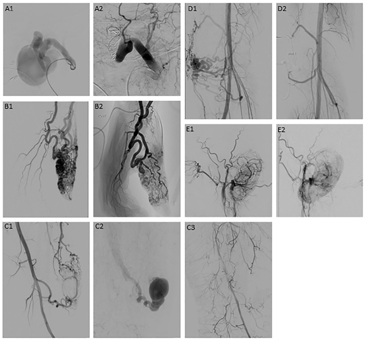
Figure 3.
Examples of cases of AVM embolization [54]; (A1,A2) AVM type I in the face ((A1) before embolization, (A2) total occlusion after embolization); (B1,B2) AVM type II in the thumb ((B1) before embolization, (B2) partial occlusion after embolization); (C1–C3) AVM type IIIa in the lower extremity ((C1) before embolization, (C2) venous outflow before embolization, (C3) total occlusion after embolization); (D1,D2) AVM type IIIb in the knee (D1) before embolization, (D2) total occlusion after embolization); (E1,E2) AVM type IV in the ear (E1) before embolization, (E2) near-total occlusion after embolization).

