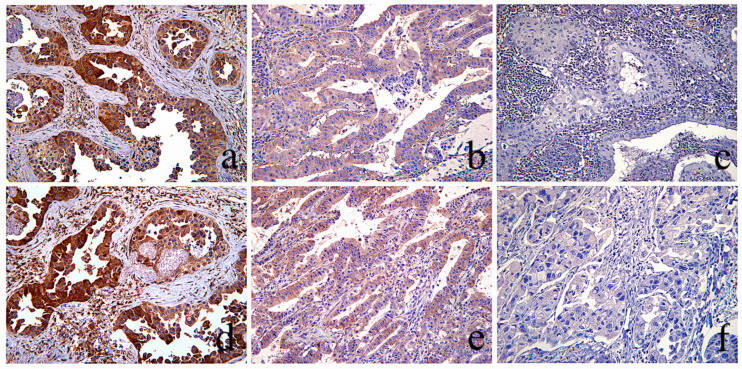
Figure 2.
LKB1 and p-AMPK immunohistochemical evaluation. Top row: Three different NSCLC tumors immunohistochemically stained for LKB1. Bottom row: Three different NSCLC tumors immunohistochemically stained against p-AMPK. (a,d) The same area of a LUAC stained with LKB1 (a), and pAMPK (b) showing cytoplasmic staining of similar, moderate and strong (scores: 2–3), intensity, therefore evaluated as “intact”. (b,e) Same area of a LUAC showing similar, very low (score: 1), staining intensity of LKB1 and pAMPK expression, evaluated as “intact”. (c,f) The same area of a LSCC with complete absence, (score: 0), of staining for both LKB1 and pAMPK was evaluated as “lost”(score: 0); [(a–e) magnification ×100, (f) magnification ×200].

