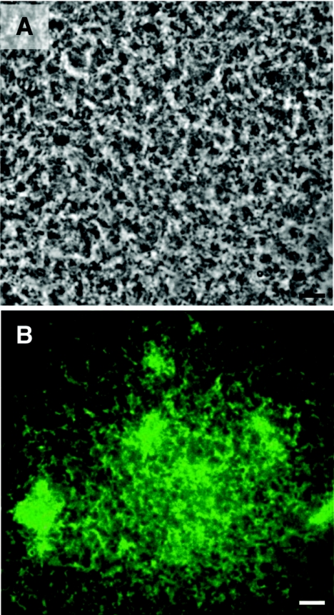
FIG. 3.
Mixed biofilms of WT and TCP− mutant strains. (A) Phase-contrast micrograph showing the differentiated biofilms developed on the surface of a squid pen by a 50:50-mixed culture of a WT and a gfp-tagged TCP− mutant strain over a 96-h period. (B) Fluorescence micrograph placed over the micrograph in panel A showing the uniform distribution of the gfp-tagged TCP− mutant strain within the mixed biofilms. Bars, 10 μm.

