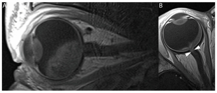
Figure 3.
Baseline and posttreatment T1-weighted contrast-enhanced MRI of a unilateral retinoblastoma patient. (A) Sagittal contrast-enhanced T1-weighted MR image of a 13-month-old patient with unilateral retinoblastoma. This pretreatment image does not show an increased enhancement of the distal optic nerve. (B) Axial contrast-enhanced T1-weighted MR image of the same patient performed after the last SIAC course (21 months of age). The patient received a total of six cycles of combined 24 mg melphalan (4 mg per cycle) and 6 mg of topotecan (1 mg per cycle). Increased contrast enhancement can be seen in the most distal part of the optic nerve (white arrowheads). After enucleation, tumor invasion was ruled out, suggesting that the increased enhancement of the optic nerve was most likely caused by an SIAC-induced inflammatory event.

