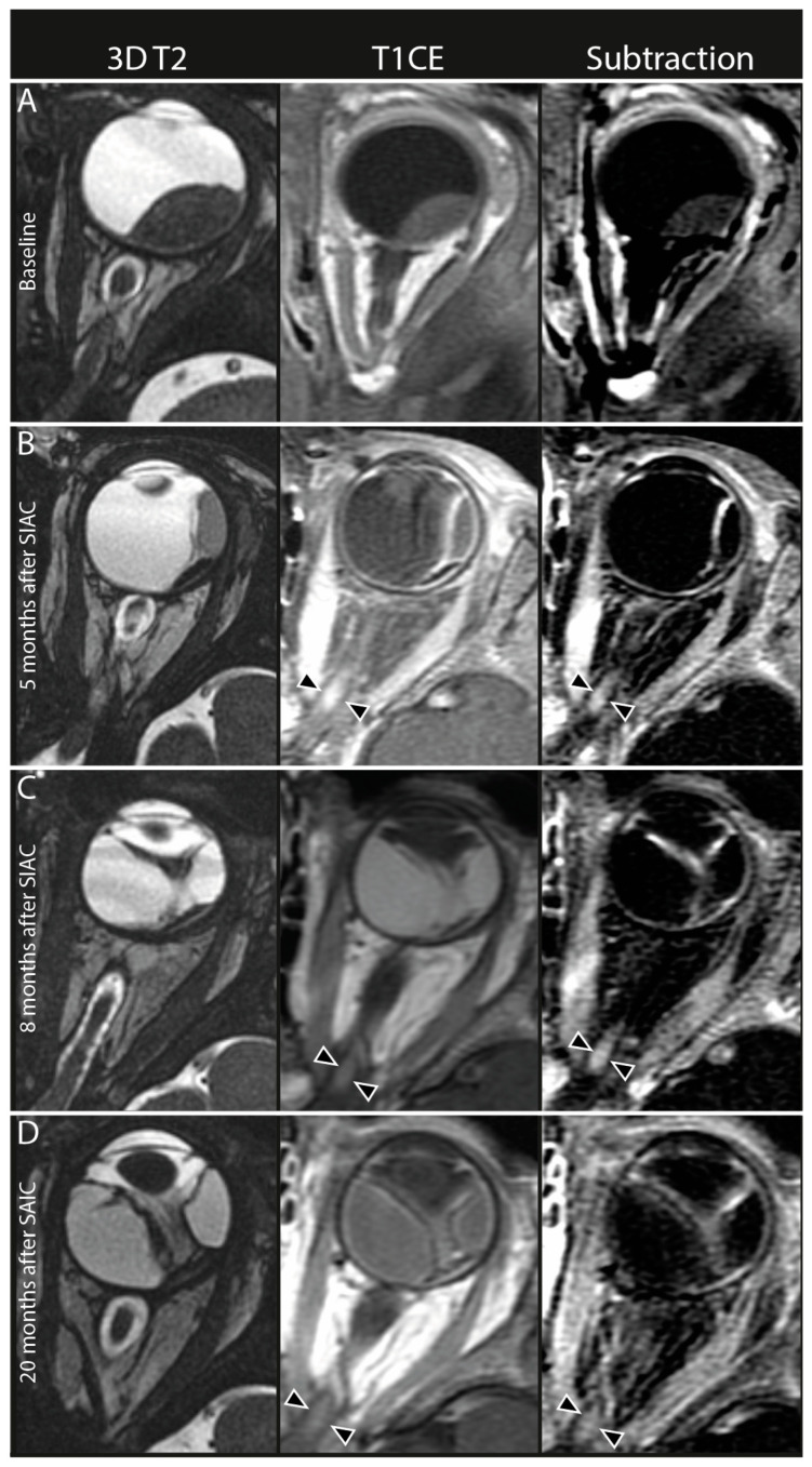
Figure 4.
Persistent optic nerve enhancement after selective intra-arterial chemotherapy (SIAC). In the left column, axial 3D T2-weighted MR images (3D T2), middle column axial contrast-enhanced T1-weighted MR images (T1CE), and in the right column axial subtraction images from contrast-enhanced T1-weighted MR images minus native T1-weighted MR images (subtraction). (A) Pretreatment MRI scan showed no optic nerve enhancement in any part of the optic nerve. (B) First follow-up MRI 5 months after the last SIAC cycle (a total of three cycles of combined 10 mg melphalan [3 mg for the first and second cycle, and 4 mg for the last cycle]. The procedure was conducted through the carotid artery with the catheter tip in the ostium of the ophthalmic artery with no wedging or complications. Enhancement of the proximal part of the optic nerve (black arrowheads). Persistent enhancement of the optic nerve 8 (C) and 20 (D) months after the last SIAC cycle (black arrowheads).

