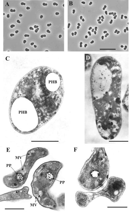
FIG. 2.
Phase-contrast micrographs of B. mobilis grown on glucose (A) and on methanol (B) in nitrogen-free medium for 6 days. Bar, 10 μm. Electron micrographs of ultrathin sections of cells of B. mobilis grown on glucose (C and E) and on methanol (D and F) in nitrogen-sufficient (C and D) or nitrogen-free (E and F) medium. MV, membrane vesicles; PHB, poly-β-hydroxybutyrate; PP, granules of polyphosphates. Bars, 0.5 μm.

