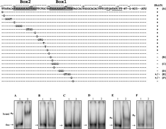
FIG. 5.
Substitution analysis of the metB2 promoter region. Upper panel: sequence of metB2 wild-type and mutated promoter region. Position +1 corresponds to the experimental transcriptional start. The deduced extended −10 box is underlined, and putative FhuR-DNA-binding box 1 and box 2 are shaded. Shifts + and − indicate the observation of a band shift or not, respectively, of the corresponding DNA fragment in the presence of the purified FhuR protein. Letters A to F on the right refer to the corresponding lower panel. Lower panels: gel mobility shift assay for FhuR binding to metB2 wild-type and mutated promoter regions. The corresponding fragments were PCR amplified and labeled. DNA probes (10 × 10−15 mole) were incubated without (lane 1) or with 3.6 μM FhuR (lane 2) in the presence of 60 mM OAS and analyzed by nondenaturing polyacrylamide gel electrophoresis. (A) metB2 wild-type promoter region. (B to F) Mutated sequences indicated on the right column of the upper panel. In each binding assay, the bands for the probe (free) and the FhuR-probe complex (bound) are indicated by an arrow and an arrow with an asterisk, respectively.

