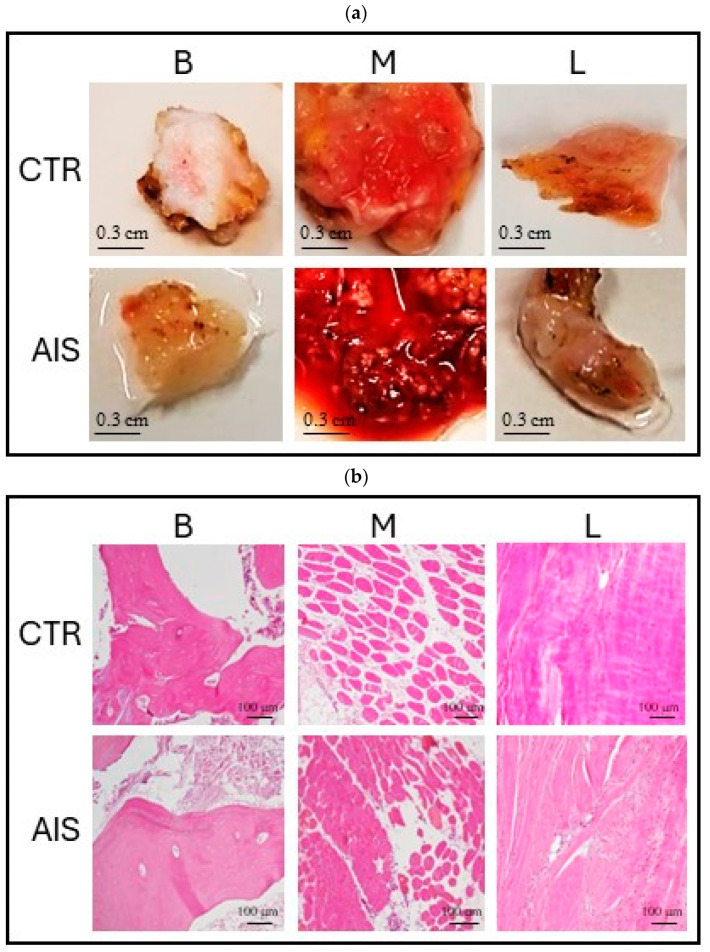
Figure 2.
Bone, muscle, and ligament tissue samples. (a) Macroscopic images of bone, muscle, and ligament samples from the control donor (top) and a representative AIS donor (bottom); (b) hematoxylin–eosin staining of bone, muscle, and ligament samples from the control donor (top) and a representative AIS donor (bottom). B = bone (spinal facet); M = paravertebral muscle; L = spinal ligament; CTR = control; AIS = adolescent idiopathic scoliosis.

