Figure 5.
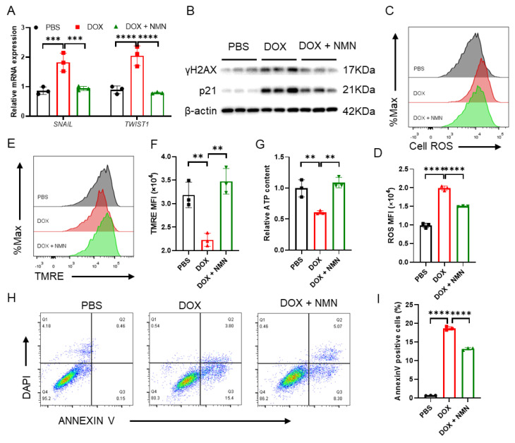
Boosting NAD+ attenuates doxorubicin-induced cellular damage in fibroblast MRC5 cells. (A) Relative mRNA expression of SNAIL and TWIST1 after treatment in human fibroblast MRC5 cells (n = 3 per group), where β-actin is used as an endogenous control. (B) Western blot of p21 and γH2AX in MRC5 cells treated with PBS, DOX or DOX + NMN (n = 3 per group), where β-actin is used as a loading control. (C) Cell ROS measurements of PBS-, DOX- or DOX + NMN-treated MRC5 cells by FACS (n = 3 per group). (D) Quantitative analysis of the mean fluorescence intensity of cell ROS (n = 3). (E) Mitochondrial membrane potential tested by FACS (n = 3 per group). (F) Quantitative analysis of the mean fluorescence intensity of mitochondrial membrane potential (n = 3 per group). (G) Cellular ATP content in MRC5 cells (n = 3 per group). (H,I) Dot plots of annexin V-FITC and 4,6-diamidino-2-phenylindole (DAPI) staining (H) and the percentages of annexin V-positive cells (I) (n = 3 per group). Data are shown as means ± standard deviations. Differences of three groups were assessed by one-way ANOVA test (A,D,F,G,I). ns, not significant, ** p < 0.01, *** p < 0.001, **** p < 0.0001.

