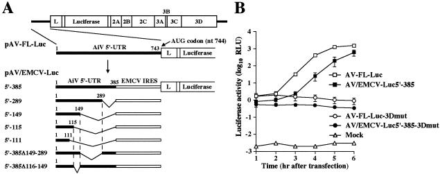
FIG. 4.
(A) Schematic diagram of the 5′ UTR of Aichi virus and chimera replicons. The thick lines indicate the Aichi virus sequences in the 5′ UTR. Each plasmid name shows the number of Aichi virus nucleotides retained at the 5′ end. The open boxes indicate EMCV IRES. (B) Luciferase activity in the transfected cells with AV-FL-Luc, AV/EMCV-Luc5′-385, AV-FL-Luc-3Dmut, and AV/EMCV-Luc5′-385-3Dmut. Vero cells were electroporated with the RNAs, and then at the indicated times after electroporation, cell lysates were prepared and analyzed for luciferase activity. The experiment was repeated at least three times. Standard deviation bars are shown.

