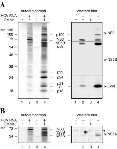
FIG. 2.
Detection of HCV antigens in HCV RNA-programmed Krebs-2 S10. (A) The assays were performed in the absence (−; lanes 1 and 3) or presence (+; lanes 2 and 4) of HCV RNA and in the absence (lanes 1 and 2) or presence (lanes 3 and 4) of CMMs as described in the legend to Fig. 1. Translation products were separated by SDS-15% PAGE and transferred onto a nitrocellulose membrane. The autoradiograph of the membrane (left) and Western blots of NS3, NS5B, and the core protein (C) (right) are shown. For Western blotting, the membrane was first probed with anti-NS3 and then sequentially stripped and reprobed with anti-NS5B and anti-Core. (B) Detection of NS5A. HCV RNA translation and separation of HCV-specific proteins by PAGE were as described for panel A. After transfer to nitrocellulose and autoradiography, the blots were reacted with an HCV NS5A-specific rabbit antiserum. The autoradiograph of the middle portion of the membrane and Western blot are shown on the left and right, respectively. An arrowhead indicates a nonspecific band. The positions of the prestained molecular weight protein markers are shown at the left.

