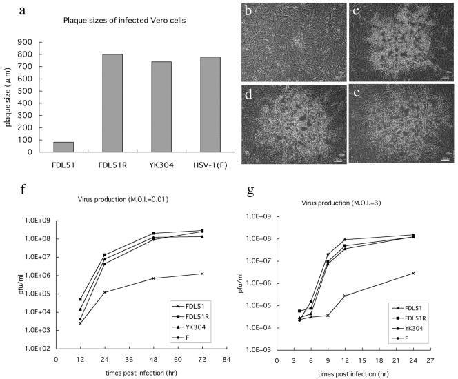
FIG. 5.
Growth properties of FDL51 in Vero cells. (a) Plaque formation of FDL51 mutant virus. Vero cells were infected with FDL51, FDL51R, YK304, or HSV-1(F) and were incubated in a medium containing gamma globulin for 48 h. Viral plaques were photographed at 48 h postinfection, and the mean diameters of 20 single plaques per virus mutant were determined as described in Materials and Methods. (b to e) Photos of viral plaques. Vero cells were infected with FDL51 (b), FDL51R (c), YK304 (d), and HSV-1(F) (e). Bars, 100 nm in all panels. (f and g) Growth kinetics of FDL51. Vero cells were infected with FDL51, FDL51R, YK304, or HSV-1(F) at an MOI of 0.01 (f) or 3 (g). At each time point, cells and supernatants were harvested and progeny virus titers were determined by plaque assays, as described in Materials and Methods. The average results of two independent experiments are shown.

