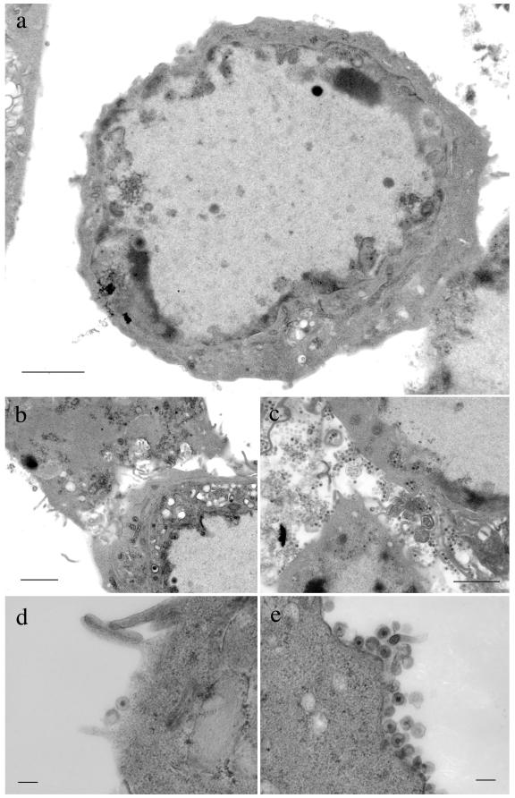
FIG. 6.
TEM analysis of extracellular regions of FDL51-infected cells. Vero cells were infected at an MOI of 3 with FDL51 (a, b, and d) or HSV-1(F) (c and e) and analyzed by electron microscopy 24 h postinfection. In FDL51-infected cells, extracellular mature virions were rarely observed in cell-to-cell junctions (b) or on the surfaces of infected cells (a and d), in contrast to wild-type HSV-1(F)-infected cells (c and e). Bars, 2 μm (a to c) or 0.2 μm (d and e).

